From glass to cloud: welcome to digital pathology
Sponsored by BarcoFrom microscopes to monitors, pathology is undergoing a digital revolution. Learn how this transformation is reshaping healthcare – and why it matters to you!

The study of the human body has fascinated us for thousands of years. From ancient civilisations to Renaissance thinkers, and from early microscopes to modern laboratories, our understanding of disease has evolved dramatically. One of the critical fields in this journey is pathology – the science of diagnosing disease by examining tissues and cells.
Today, pathology is entering a new era: the digital age. Known as digital pathology, this innovation is transforming how pathologists work, making diagnoses faster, with high precision, and enabling easy collaboration with other specialists around the globe.
What is digital pathology?
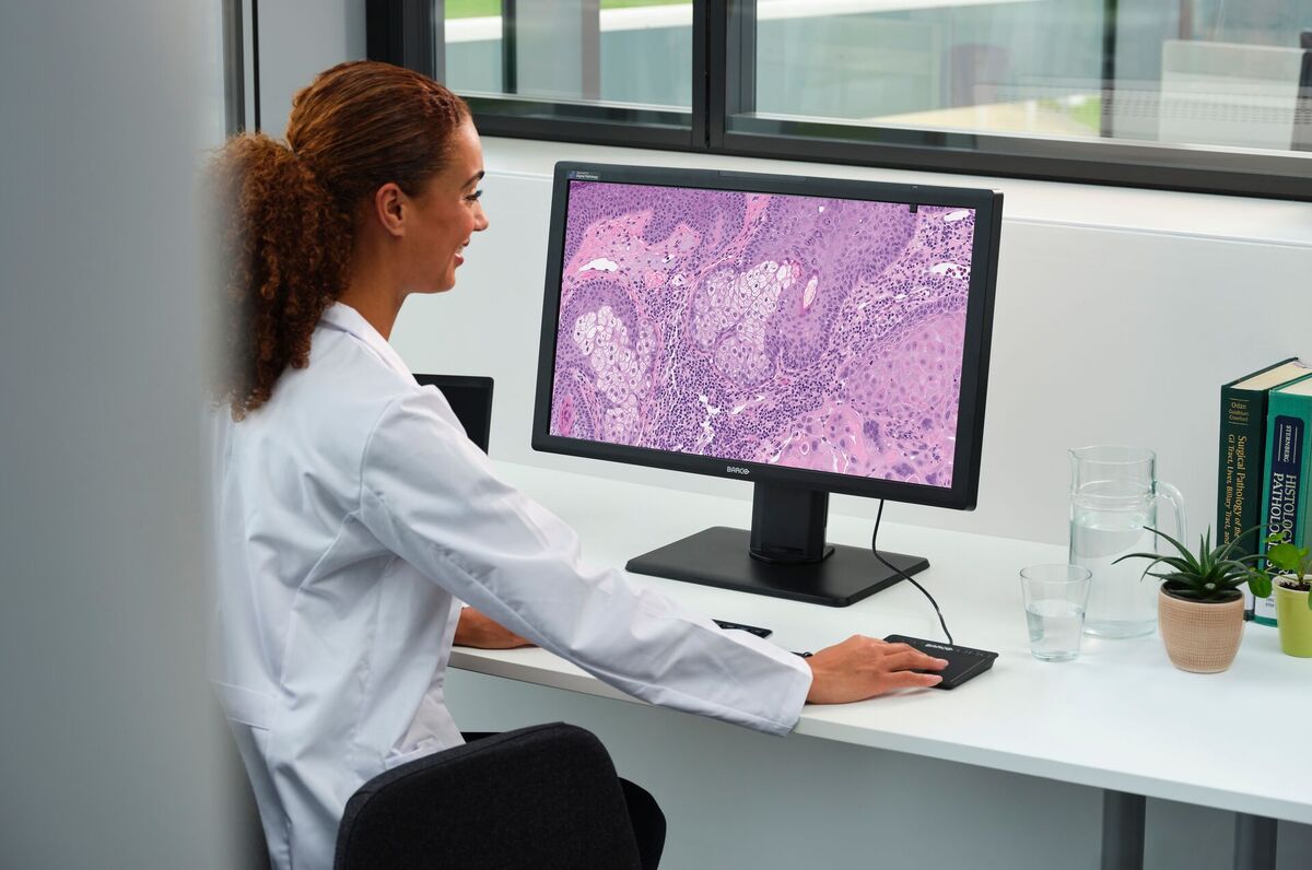
At its core, digital pathology is the process of digitising traditional glass slides – those thin slices of tissue you might imagine under a microscope – as high-resolution digital images. These images can then be viewed, analysed and shared using specialised software and medical displays.
Instead of peering through a microscope, pathologists can now examine tissue samples on a computer screen, zooming in and out, adding notes and even collaborating with colleagues across the globe in real time. On top of that, digitised images allow AI algorithms to run, supporting pathologists as they find and identify diseases.
Why now?
Although the idea of virtual microscopy has been around since the 1990s, the technology needed to make it practical – such as powerful scanners, fast internet and secure digital storage – has only recently become widely available.
But technology isn’t the only driver. The world’s population is aging, and diseases such as cancer are becoming more common. Diagnostic processes are also becoming more advanced and personalised, requiring healthcare professionals to spend more time on them. At the same time, there’s a growing shortage of pathologists, especially as many experienced professionals retire. This means more work for fewer people – and a greater need for efficient, flexible solutions.
Digital pathology helps meet this demand by speeding up workflows, reducing the need to physically ship slides for second opinions, and even allowing pathologists to work from home.
How it works
A digital pathology system includes several key components:
Slide scanner. This device scans glass slides and creates ultra-high-resolution digital images – some as large as 10Gb, the equivalent of 2,000 high-quality photos.
Viewing software. Once scanned, the images are compressed and optimised for viewing. Pathologists can then analyse them on a display, just as they would with a microscope.
Display. High-quality monitors ensure that every detail of the tissue is visible and accurate.
Storage. Digital slides are stored in secure systems, either on-site or in the cloud, where they can be accessed anytime.
Benefits that matter
Digital pathology isn’t just a tech upgrade – it brings real-world benefits that impact patients, doctors, and healthcare systems alike.
Faster diagnoses
Digital slides can be shared instantly, eliminating the delays caused by shipping glass slides. This means quicker diagnoses and faster treatment decisions.
Remote collaboration
Pathologists can consult with specialists anywhere in the world, viewing the same slide at the same time. This is especially valuable in rural or underserved areas.
Better documentation
Digital slides can be annotated without altering the original image. These notes help with teaching, research and future case reviews.
AI and big data
With thousands of digital slides stored, artificial intelligence (AI) can be trained to recognise patterns, highlight abnormalities and even predict disease outcomes. These tools act like a second pair of eyes, supporting pathologists in their work.
Challenges to overcome
Of course, going digital isn’t without its hurdles. Setting up a digital pathology system requires a significant investment in equipment, software and training. There are also technical challenges, such as ensuring fast and secure data storage and meeting strict regulatory standards.
But for many institutions, the long-term benefits – greater efficiency, improved accuracy and future-proofing – far outweigh the initial costs.
Why it matters to you
Even if you’ve never heard of pathology before, it plays a vital role in your healthcare. Every time a tissue biopsy is taken – whether it’s a mole, a lump or a suspicious spot – it’s a pathologist who examines it and helps determine what’s next.
By making this process faster, more accurate and more collaborative, digital pathology is helping ensure better and faster results for patients everywhere.
A glimpse into the future
Imagine a world where a pathologist in the United Kingdom can instantly consult with a specialist in Japan. Where AI tools help detect cancer earlier and more accurately. Where medical students learn from real cases using interactive digital slides. This is the promise of digital pathology.
Digital pathology is more than just a technological upgrade – it’s a revolution in how we understand and diagnose disease. It empowers pathologists, supports healthcare systems and, most importantly, benefits patients.
by Mirelva Drost, VP Product Management for Diagnostic Imaging, Barco

Business Reporter Team
You may also like
Related Articles
Most Viewed
Winston House, 3rd Floor, Units 306-309, 2-4 Dollis Park, London, N3 1HF
23-29 Hendon Lane, London, N3 1RT
020 8349 4363
© 2025, Lyonsdown Limited. Business Reporter® is a registered trademark of Lyonsdown Ltd. VAT registration number: 830519543


